R2 or Not? September 2018
Total: 25 responses
Image 1
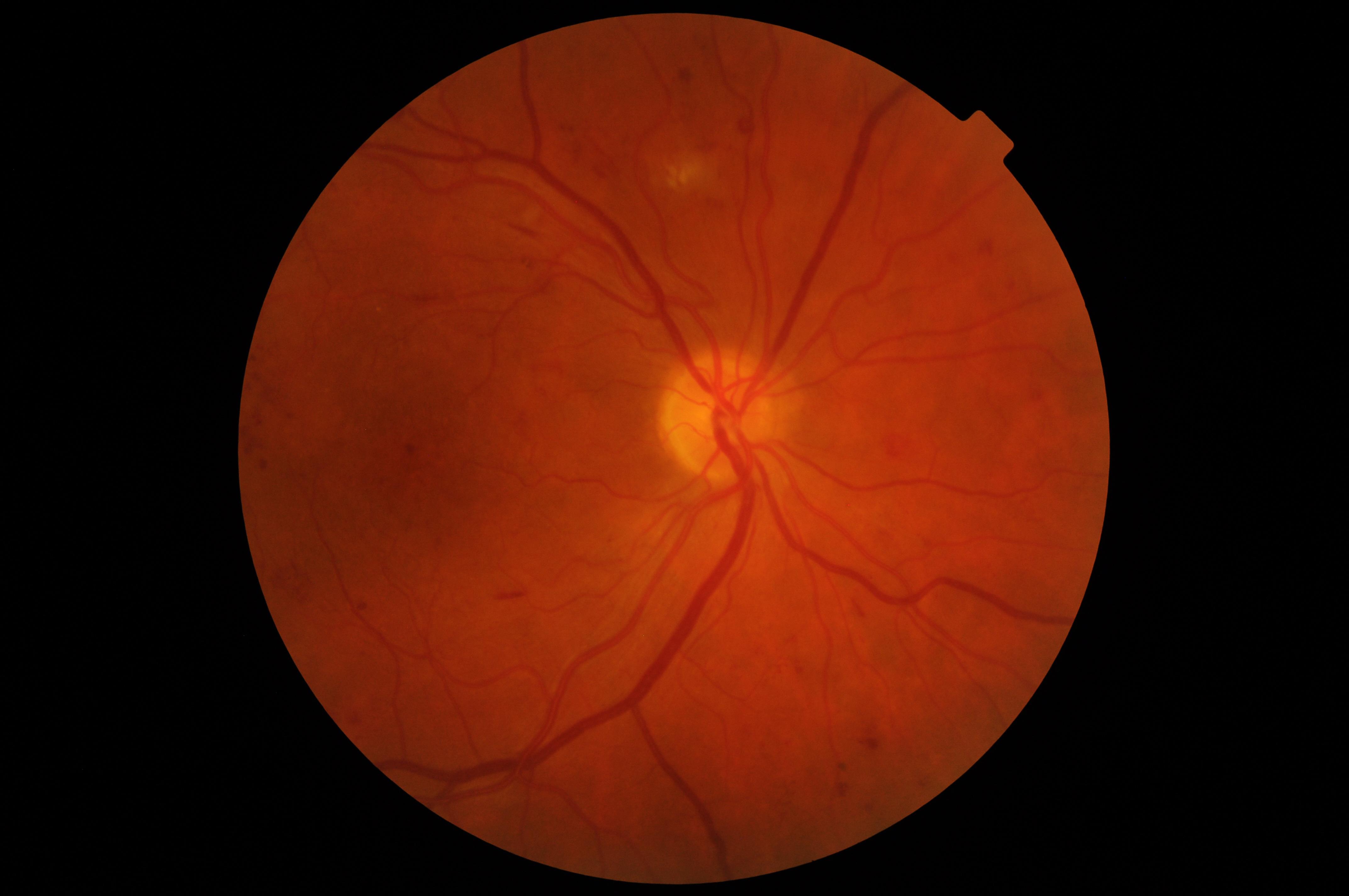
Overall Result:
R2: 60%
Not R2: 40%
Broken down by grader level:
Trainee Graders (6)
Qualified Graders (13)
ROG/L3 Graders (6)
Comments from those who said R2:
- Borderline assessable (ROG)
- suspect IRMA 9/10 o clock
- Minimum R2
- Seems like a comfortable R2 on MBH alone - even without having to see the macula view you've got over 8 MBH and some suitable other aggregate features (ROG)
Comments from those who said NOT R2:
Image 2
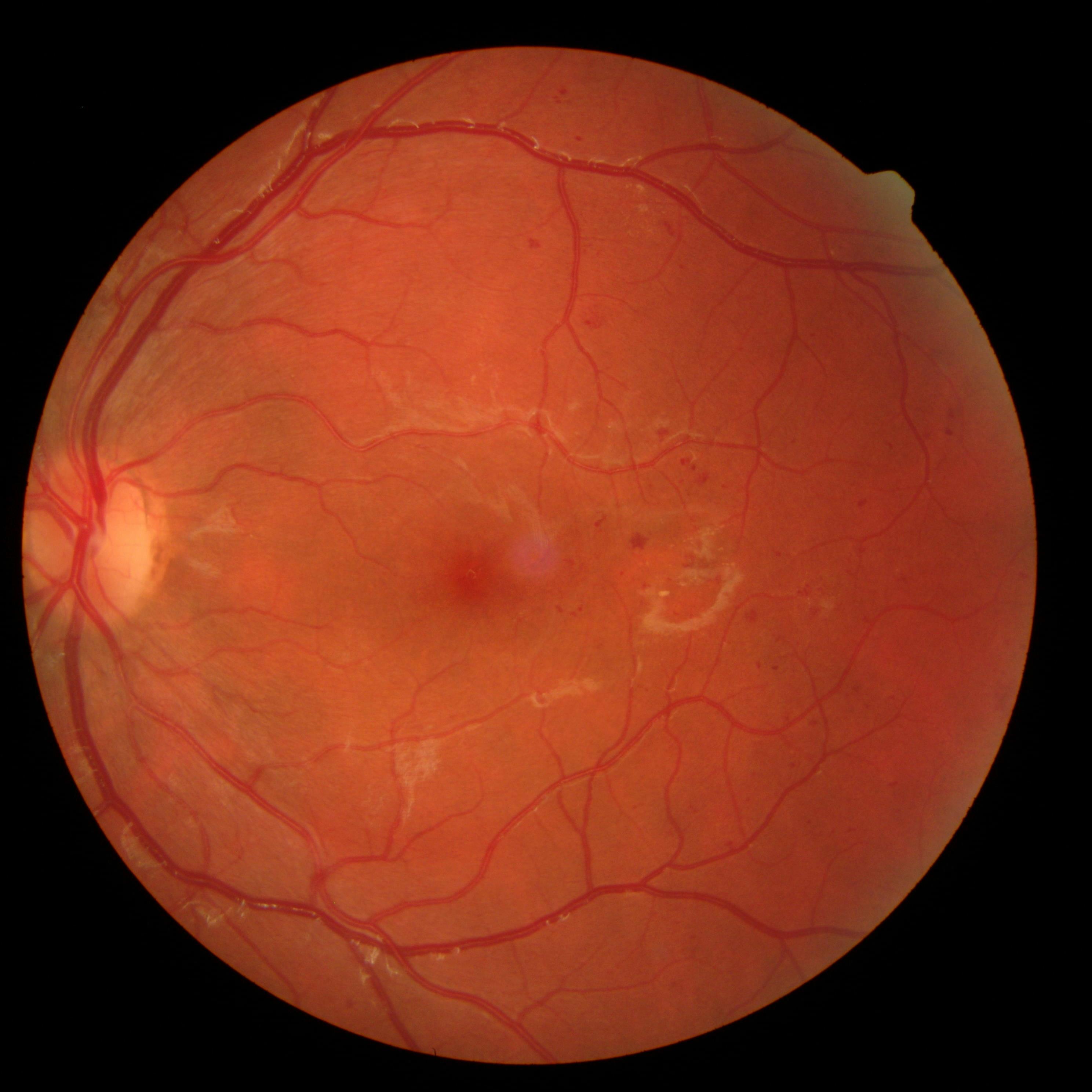
Overall Result:
R2: 44%
Not R2: 66%
Broken down by grader level:
Trainee Graders (6)
Qualified Graders (13)
ROG/L3 Graders (6)
Comments from those who said R2:
- not referrable - but enough haemms and MA
Comments from those who said NOT R2:
- Potentially IRMA but black and white images reveal unlikely, though referrable as M1
- Looking under on MBH and without any strong suspicion of IRMA or other vascular abnormalities. What makes this an even more comfortable R1 is the faint M1 marker (within 0.5DD inferior temporal) that would probably get this into ophthalmology anyway
Image 3
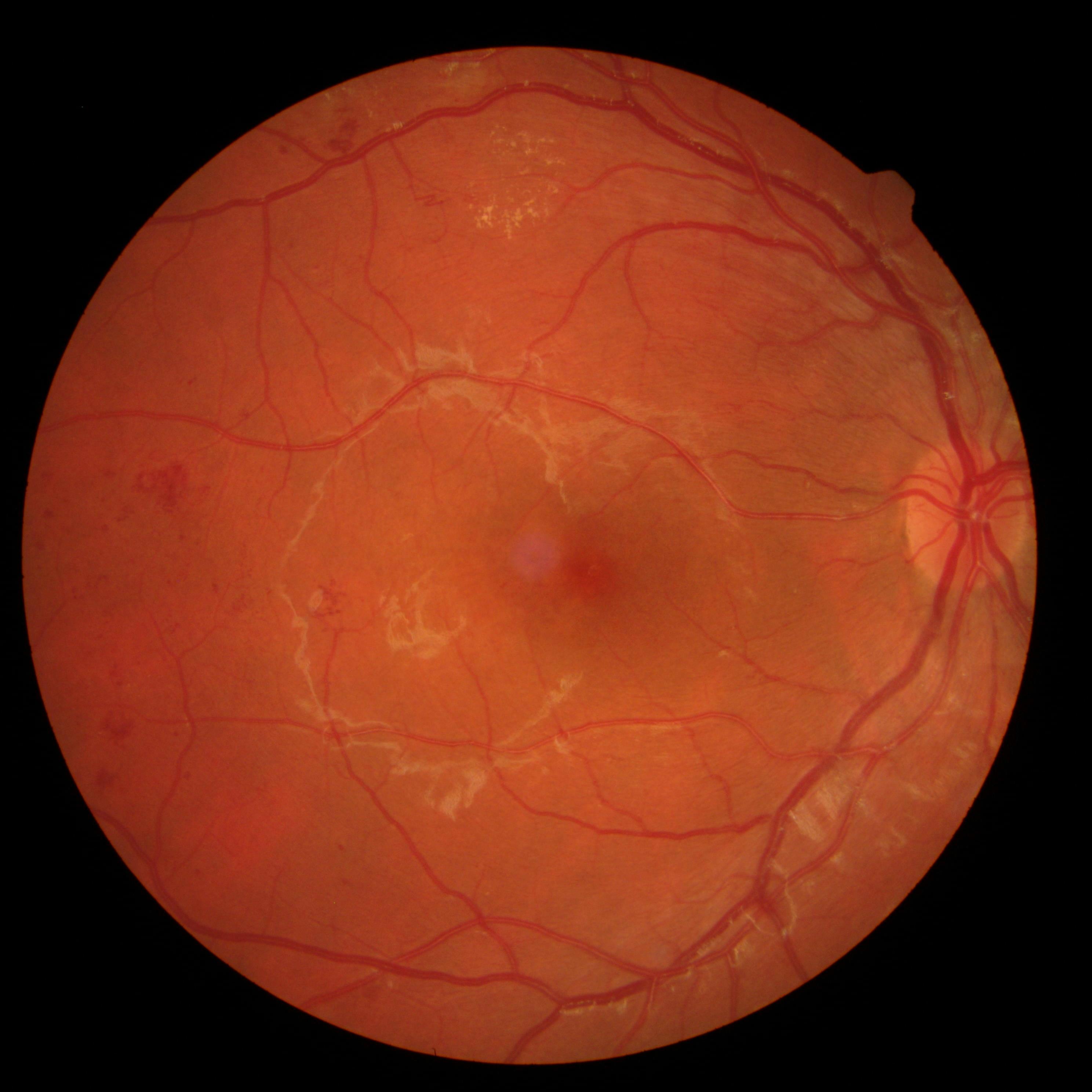
Overall Result:
R2: 80%
Not R2: 20%
Broken down by grader level:
Trainee Graders (6)
Qualified Graders (13)
ROG/L3 Graders (6)
Comments from those who said R2:
- odd vessel structure 11 o clock
- Minimum R2, possible early NVE
Comments from those who said NOT R2:
- There looks to be a loop 3DD from fovea at 11 o'clock and the MBH is on the low side so a solid R1 that may find it's way into the DS pathway for a little closer observation. It seems a little strange to me that we are potentially grading R1 or R2 on an MBH count using just 1 view - one view is obviously good for a borderline vascular abnormality but seems like an unusual situation for weighing MBH as I'm not sure users will know consistently whether assume a similar MBH spread across the missing view or to assume there is nothing outside of this view (which would be weird). (ROG)