R2 or Not? July 2018
Total: 37 responses
Image 1
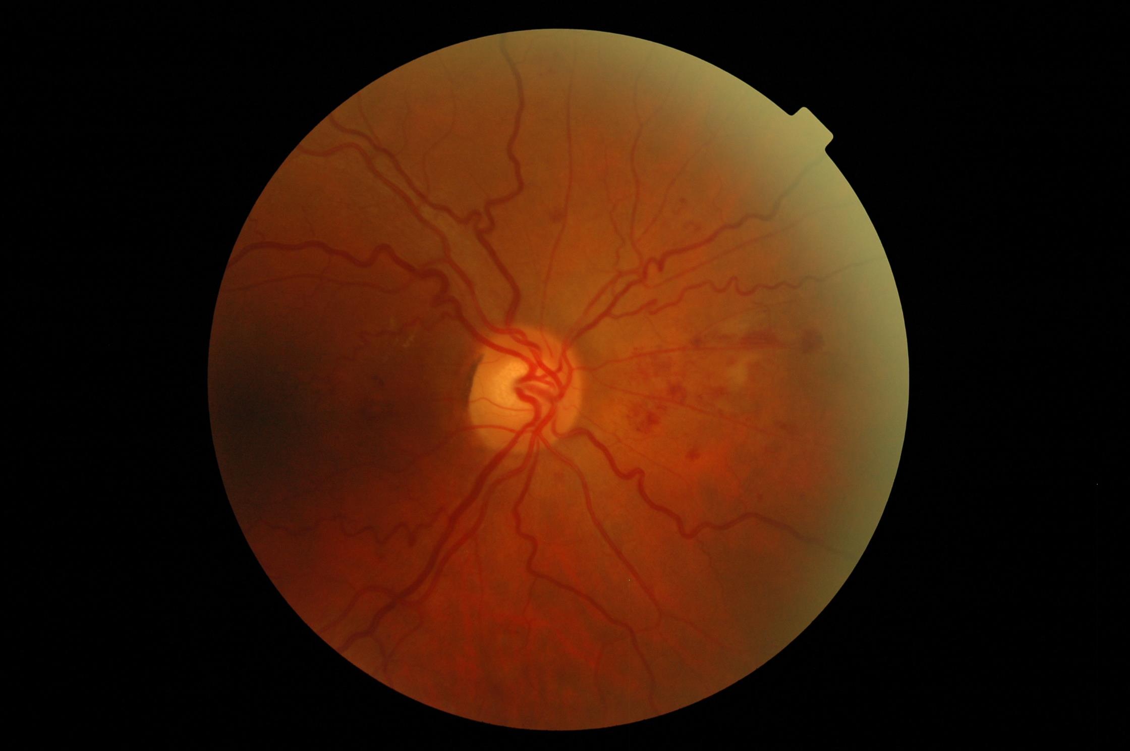
Overall Result:
R2: 68%
Not R2: 32%
Broken down by grader level:
Trainee Graders (6)
Qualified Graders (21)
ROG/L3 Graders (10)
Comments from those who said R2:
- Dark image difficult to be sure how much DR in macular area.
- 1st image considering as R2 due to the second image being from the same patient so strangely I'm combining features from question 1&2 - it's possible that these were taken at separate encounters so not viable to weigh them together but it's more fun to combine them
Comments from those who said NOT R2:
- Just from nasal image, would not constitute MBH and no other distinguishable R2 features (ROG)
- Suspect NVE present so safer R3
- Very difficult to judge from only nasal view, I would say R1 and half as unable to see macular view. (ROG)
- Not enough separate larger haemorrhages? (ROG)
- just from the nasal images it is difficult to assess, if the macula images is quiet the definitely R1 otherwise I would possibly give it the benefit of doubt (ROG)
Image 2
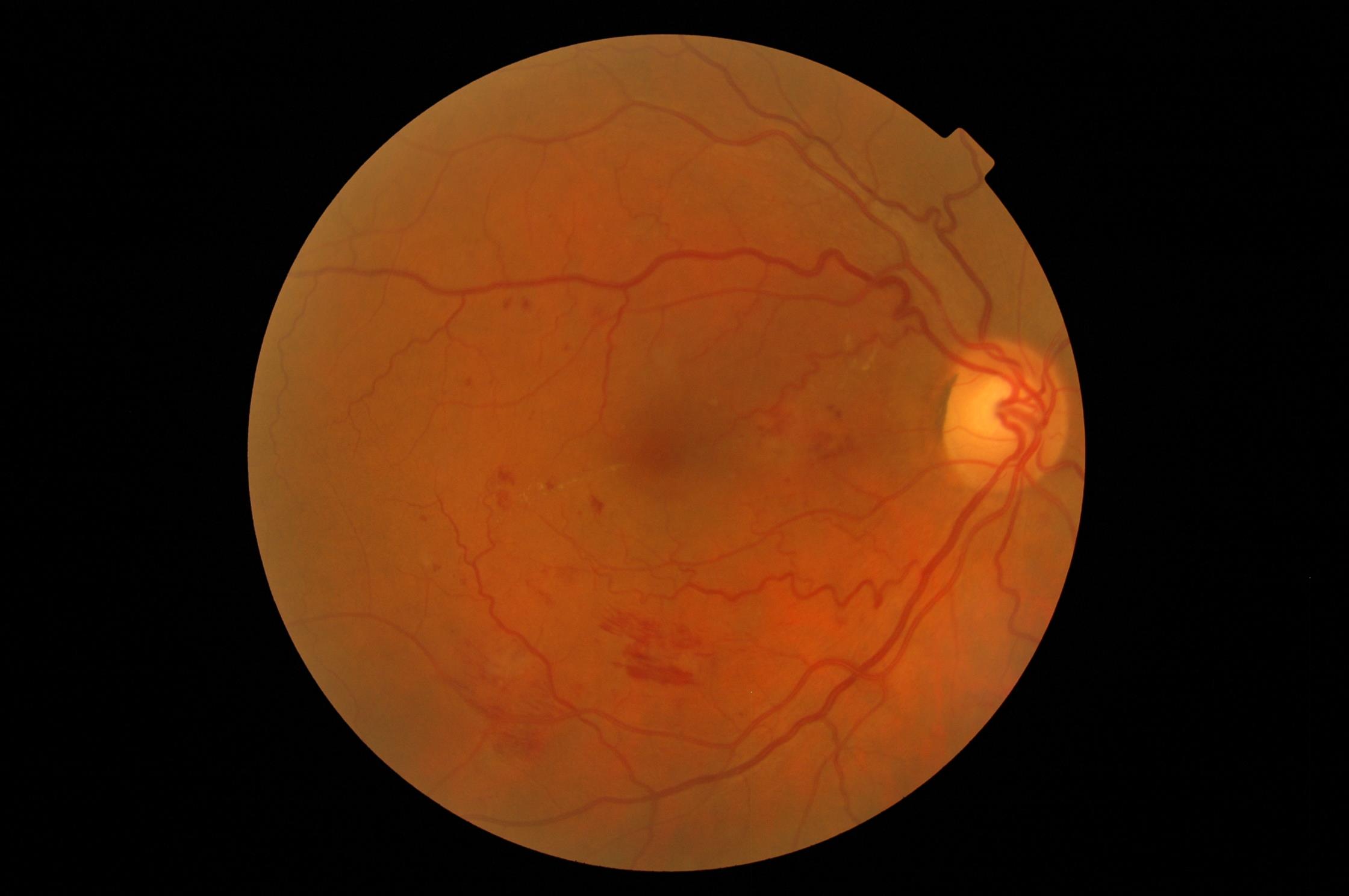
Overall Result:
R2: 85%
Not R2: 15%
Broken down by grader level:
Trainee Graders (6)
Qualified Graders (21)
ROG/L3 Graders (10)
Comments from those who said R2:
- R1 and half as unable to see nasal view. If nasal view is clear, then it is R1.
- definitely busy enough just in the macula view to be R2
- When put together with Image 1 (which is the same eye), I would put it as R2, because there are just enough big haemorrhages, and I wouldn't want to put this as Annual Recall (although I would consider putting this as R1 with BRVO).
Comments from those who said NOT R2:
- Again, doesn't quite meet MBH, would be a good candidate for 3-month DS (if exudate wasn't there) however, referable due to exudate (ROG)
- Don't think R2. Very questionable if it is. Big haemorrhage but as picture is slightly hazy it has probably been assessed as R2 to be viewed in SLB.
- Evidence of NVE/R3
Image 3
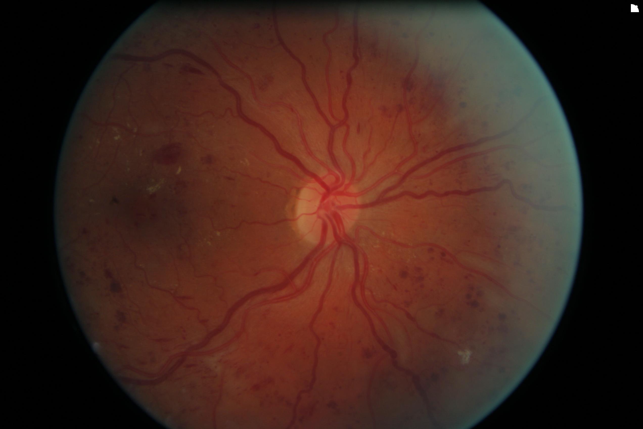
Overall Result:
R2: 86%
Not R2: 14%
Broken down by grader level:
Trainee Graders (6)
Qualified Graders (21)
ROG/L3 Graders (10)
Comments from those who said R2:
- A very good example of R2, IRMA, MBH, beading. Given fibrosis in bottom right, prudent to look for NVE in this area, some of the vessels here do look like NVE. (ROG)
- Also M1, may be CRVO.
- Borderline R2/3, but cannot clearly see any R3 markers.
- the images of this px need careful examination, very busy and there could be NVE but from just the one image it is possible a very sever R2
- i would be looking very carefully for R3 changes
Comments from those who said NOT R2:
- This is R3 as there are new vessels 2-3DD inferior nasal to disc - it's a pretty torn up area as there's a lot of venous beading in that region (anecdotally for me VB is the most severe R2 lesion prognostically as it speaks of vast structural loss due to DR) and a small patch of fibrosis (ROG)
- MBH 4, inferiorly white are ? fibrosis, some vessels near area of venous beading - ? IRMA ? area of previous focal ischaemia with NVS and fibrosis. Picture is of severe R2 changes, indicative of severe ischaemia. High risk Ht, renal failure, poor control . Very high risk need urgent referral whatever the grade. (ROG)